How Tooth Decay Really Progresses: From Early Enamel Changes to a Cavity

Written by: Skylar Sun
A cavity is easy to picture: a hole in a tooth.
The more useful idea is that the hole is not the beginning of tooth decay. It is a later physical result of a process that has already been happening at the tooth surface.
That distinction changes how people think about prevention.
According to the National Institute of Dental and Craniofacial Research (NIDCR), early tooth decay can begin with mineral loss from enamel before an actual cavity has formed.
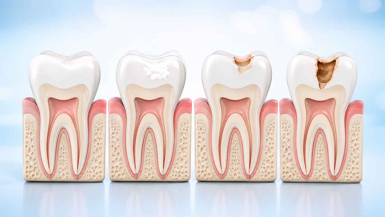
Repeated acid exposure can cause enamel to lose minerals, and a white spot may sometimes appear where that mineral loss has occurred. At this early stage, the process may sometimes be stopped or reversed because minerals from saliva and fluoride can support remineralization.
Once enough tooth structure has broken down to form a cavity, however, the physical damage is permanent and generally requires professional dental treatment. NIDCR explains that a cavity represents permanent structural damage that a dentist typically repairs after removing decayed tissue.
So the important dividing line is not simply:
“Do I have tooth decay?”
It is:
“Has the process remained at an early mineral-loss stage, or has tooth structure already broken down?”
You cannot reliably answer that question from a bathroom mirror. Understanding how the process develops, however, can make dental recommendations much easier to interpret.
A Tooth Is Constantly Losing and Regaining Minerals
Enamel may look static, but its surface exists in a changing chemical environment.
NIDCR describes this process almost like a daily tug-of-war. Bacteria in dental plaque can use sugars and starches from foods and drinks to produce acids. Those acids contribute to mineral loss from enamel. Saliva supplies minerals such as calcium and phosphate, while fluoride from toothpaste, water, and other sources can help replace minerals that have been lost.
This is better understood as a repeated process than as one dramatic event.
One cookie does not suddenly drill a cavity into a tooth.
One brushing session does not permanently reset every risk.
What matters is the balance that develops over time.
Stage One: Mineral Loss Without a Hole
Early decay may begin with demineralization.
A white spot can sometimes appear where minerals have been lost from enamel. NIDCR identifies this as a possible sign of early decay and explains that the process may still be stopped or reversed before the enamel surface has broken down into a cavity.
This stage is often misunderstood.
“Reversing early decay” does not mean regrowing a large missing part of a tooth.
It means shifting the mineral balance while the enamel surface has not yet physically collapsed into a cavity.
That distinction is important because remineralization and restoration describe two different situations.
Stage Two: Enamel Breakdown and Cavity Formation
If mineral loss continues faster than mineral replacement, enamel can become progressively weakened.
Eventually, the structure may break down and form a cavity.
At that point, ordinary fluoride toothpaste cannot reconstruct the missing anatomy.
Fluoride remains important for protecting surrounding tooth surfaces and helping prevent further demineralization, but it should not be interpreted as a way to “grow back” a physical hole.
NIDCR distinguishes clearly between early mineral loss that may be reversible and a cavity, which represents permanent damage that requires dental repair.
Stage Three: Decay Can Move Deeper
A tooth is not made entirely of enamel.
Beneath enamel is dentin, and deeper inside is the pulp, which contains nerves, blood vessels, and connective tissue.
As decay becomes more advanced, symptoms may become more likely.
NIDCR notes that early tooth decay usually has no symptoms. As decay progresses, a person may develop toothache or sensitivity to sweets, hot foods, or cold foods. If infection develops, additional symptoms can include pain, facial swelling, or fever.
The important wording is may.
Pain intensity does not tell you the exact depth of decay.
Some teeth with significant decay can produce surprisingly little discomfort. Other painful teeth may hurt for reasons unrelated to cavities.
Understanding the Tooth Decay Progression Map
The following map is designed to make the stages easier to understand.
| Conceptual stage | What may be happening | Can you confirm it yourself? | Typical professional question |
|---|---|---|---|
| Mineral balance | Enamel losing and regaining minerals | No | Is this surface healthy or demineralized? |
| Early demineralization | Mineral loss without a cavity | No | Can this area be stabilized or remineralized? |
| Cavitation | Tooth surface has physically broken down | No | How much tooth structure is affected? |
| Deeper decay | Decay extends farther into the tooth | No | How close is the lesion to the pulp? |
| Pulp or infection concern | Inner tooth tissues may be involved | No | What treatment is appropriate for the actual diagnosis? |
The map is best used as an explanation of the process, not as a way to assign your own tooth to a stage.
A dentist may need visual examination, tactile findings, dental history, symptoms, and imaging when appropriate to determine what is actually happening.
Why Sugar Frequency Matters
People often focus only on how many grams of sugar they consume.
For tooth decay, frequency of exposure can also matter.
NIDCR explains that when decay-related bacteria come into contact with sugars and starches, they can produce acids that attack enamel. If this happens repeatedly throughout the day, the teeth can experience repeated acid challenges before enough mineral recovery has occurred.
That means a sweet drink consumed during a meal can create a different exposure pattern from the same drink being sipped slowly for several hours.
For dental health, “how often?” can sometimes be as useful a question as “how much?”
Starch Belongs in the Conversation Too
People commonly associate cavities only with candy.
NIDCR's explanation of the decay process includes foods and drinks containing both sugars and starches because oral bacteria can use these carbohydrates to produce acids.
That does not mean bread, fruit, milk, or every carbohydrate should be treated as a dental threat.
A useful cavity-prevention discussion looks at the whole pattern rather than labeling individual foods as automatically “good” or “bad.”
Risk can be influenced by a combination of:
- bacterial plaque;
- eating and drinking patterns;
- exposure frequency;
- saliva;
- fluoride;
- tooth anatomy;
- existing dental restorations;
- oral hygiene;
- individual cavity history.
No single food explains every cavity.
Why Saliva Is Part of the Decay Story
Saliva helps shift the mineral balance in a protective direction.
According to NIDCR, saliva supplies minerals including calcium and phosphate that can help replace minerals lost from enamel during acid attacks.
That is one reason persistent dry mouth can matter when a dentist evaluates cavity risk.
Someone with reduced salivary protection may need a different prevention discussion from someone with normal salivary flow.
This does not mean everyone with dry mouth will develop cavities.
Saliva is one protective factor among many.
Fluoride Does More Than “Make Teeth Strong”
“Fluoride strengthens teeth” is common wording, but it does not fully explain what fluoride is doing.
NIDCR explains that fluoride can help prevent mineral loss, replace minerals in early demineralized enamel, and reduce the ability of decay-related bacteria to produce acid.
That is why fluoride can be particularly important before cavitation.
Once the tooth surface has physically broken down into a cavity, fluoride does not recreate the missing tooth structure.
This distinction helps explain why a dentist may recommend preventive management for one area while recommending restorative treatment for another.
White Spots Are Not Automatically Cavities
Because white spots can sometimes occur with early demineralization, readers may begin inspecting every pale area on their teeth.
That is not a reliable way to diagnose decay.
White areas can have multiple explanations.
A dentist may consider:
- location;
- appearance;
- surface texture;
- dental history;
- whether the area is changing;
- surrounding tooth structure;
- other clinical findings.
NIDCR does identify white spots as one possible sign of mineral loss during early decay, but appearance alone cannot determine the cause of every white area.
Do not assume that every white mark represents an active cavity.
Brown or Black Does Not Automatically Mean Decay Either
Color is similarly unreliable at the other end of the spectrum.
Teeth can collect external stain.
Natural grooves can look dark.
Old restorations can change the way surrounding tooth structure appears.
Areas that have experienced previous mineral changes may also look different from completely unaffected enamel.
NIDCR notes that dentists may observe white, brown, or black staining when evaluating tooth decay, but diagnosis can also involve examination for soft or sticky areas and dental X-rays when appropriate.
That is why:
dark spot = cavity
is too simplistic.
Color can raise a question. It cannot answer the whole question.
Why Cavities Between Teeth Can Be Hard to See
The contact areas between neighboring teeth can be difficult to inspect directly at home.
Even during a dental examination, some proximal surfaces are not fully visible.
Depending on the patient's history, cavity risk, and clinical findings, a dentist may use dental radiographs to evaluate areas that cannot be assessed adequately through direct visual examination.
NIDCR notes that X-rays can reveal decay that may not otherwise be visible during a routine examination.
This is one reason:
“I don't see a hole.”
is not a strong way to rule out decay.
Root Cavities Are Different From Enamel Cavities
The crowns of teeth are protected by enamel.
Root surfaces are different.
When gums recede, tooth roots can become exposed to the oral environment. NIDCR notes that exposed root surfaces can become vulnerable to decay-causing bacteria, particularly in people with gum recession.
A dentist who mentions “root caries” is therefore not simply describing another cavity on the chewing surface.
The location and tooth structure involved are different.
That can affect preventive and restorative decisions.
A Filling Treats Damage, Not the Entire Risk Pattern
A filling restores tooth structure after a cavity has developed.It does not permanently remove the conditions that allowed decay to occur.
A person who has had one filling can still develop new decay:
- on another tooth;
- between teeth;
- near an existing restoration;
- on an exposed root surface;
- elsewhere in the mouth.
That is why restorative treatment and prevention are connected but not identical.
The restoration repairs damaged tooth structure.
The preventive strategy addresses the conditions that may contribute to future disease.
Why a Refilled Tooth Is Not Necessarily a “Failed Filling”
Dental restorations exist in an environment that continues to change.
A tooth may require additional treatment years later because of:
- new decay;
- tooth fracture;
- restoration wear;
- changes in the surrounding tooth;
- another dental problem.
That does not automatically prove that the original restoration was poorly placed or that the filling material itself failed.
A useful question is:
“Is the problem around the existing restoration, underneath it, inside another part of the tooth, or somewhere else?”
That moves the conversation toward actual findings rather than assumptions.
The Cavity Timeline Has No Universal Clock
One of the most misleading questions online is:
“How long does it take for a cavity to form?”
There is no single countdown that applies to every tooth.
Progression can vary according to factors such as:
- the tooth surface involved;
- the person's cavity history;
- fluoride exposure;
- saliva;
- eating and drinking patterns;
- frequency of acid-producing exposures;
- oral hygiene;
- existing restorations;
- whether the lesion is active or stable.
Because of that variation, a generic rule such as “a cavity takes exactly six months” or “one year” can create false reassurance or unnecessary anxiety.
When a dentist is monitoring an area, comparison over time is more useful than a universal internet timeline.
Questions to Bring to Your Next Dental Visit
If you are trying to understand your cavity risk or a tooth that is being monitored, these questions can make the appointment more productive.
Your Recent Dental History
- Have you had a cavity in the last few years?
- Has a dentist been monitoring a specific tooth?
- Do you have several existing fillings or other restorations?
- Has persistent dry mouth become a problem?
- Has your snacking or drinking pattern changed?
Symptoms You Have Noticed
- Cold sensitivity?
- Sweet sensitivity?
- Biting discomfort?
- Spontaneous toothache?
- Food repeatedly trapping in one area?
- No symptoms at all?
Daily Exposure Patterns
- Frequent sugary drinks?
- Repeated snacking?
- Slowly sipping sweet drinks over long periods?
- Any medications that may contribute to dry mouth?
- Regular fluoride toothpaste use?
This worksheet is simply a way to organize information for a dental conversation.
It does not calculate a cavity score or determine whether treatment is necessary.
What “Watch This Area” Can Mean
Patients sometimes become anxious when a dentist says:
“We're going to watch this area.”
Monitoring does not necessarily mean ignoring decay.
Depending on what the dentist sees, an area may be early, non-cavitated, stable, or suitable for preventive management and follow-up rather than immediate restoration.
NIDCR distinguishes between early tooth decay that may potentially be stopped or reversed and a true cavity representing permanent tooth damage.
A useful follow-up question is:
“What change would make you recommend treatment instead of continued monitoring?”
That helps clarify what the dentist is watching and why.
When a Filling Becomes the Appropriate Conversation
When a true cavity has formed, the physical defect generally requires professional restoration.
NIDCR explains that dentists commonly treat cavities by removing decayed tooth tissue and restoring the missing area with a filling material.
The American Dental Association (ADA) also notes that filling-material decisions are individualized. A dentist may consider factors such as the size and location of the cavity, durability, cosmetic considerations, insurance coverage, and out-of-pocket cost when discussing options with a patient.
That means there is not one universally “best” filling material for every tooth and every person.
The choice depends on the actual clinical situation and patient priorities.
What Happens if Decay Reaches Deeper Tooth Structures?
As decay progresses farther into a tooth, the treatment discussion can become more complex.
A dentist may need to consider:
- how much healthy tooth structure remains;
- whether the pulp is involved;
- whether infection is present;
- whether the tooth can be predictably restored;
- the condition of surrounding structures.
Depending on the diagnosis, treatment may involve more than a simple filling.
This article does not attempt to determine which treatment a particular tooth requires. That decision depends on clinical examination and any additional tests or imaging the dentist considers appropriate.
Why Pain Is a Poor Screening System
Waiting until a tooth hurts is not a reliable cavity-detection strategy.
NIDCR specifically states that early tooth decay usually has no symptoms. Sensitivity or toothache may develop only as the disease progresses.
That is why the absence of pain does not prove that a tooth is free of decay.
It also explains why routine dental examinations can identify problems before discomfort becomes the first warning sign.
Preventive care and emergency care begin at very different points in the disease process.
You Cannot Determine Cavity Depth at Home
A mirror can show changes.
A phone camera can record them.
Neither can reliably determine how deeply decay extends into a tooth.
You cannot determine cavity depth at home.
That requires professional evaluation.
What you can observe includes:
- a new visible change;
- sensitivity;
- food trapping;
- discomfort when chewing;
- changes near an existing filling;
- whether a problem appears to be progressing.
Those observations are useful to report.
They should not be turned into home diagnostic tests.
What You Can Realistically Do at Home
You cannot diagnose the depth or activity of a cavity on your own, but everyday habits can influence the environment around the teeth.
General evidence-based priorities include:
- brushing with fluoride toothpaste;
- cleaning between teeth consistently;
- reducing repeated sugary or starchy exposures throughout the day;
- choosing water more often for routine hydration;
- addressing persistent dry mouth with appropriate professional guidance;
- attending dental examinations based on individual needs.
NIDCR recommends fluoride, consistent oral hygiene, limiting frequent sugary foods and drinks, and regular dental care as part of tooth-decay prevention.
These steps reduce risk; they do not guarantee that a person will never develop a cavity.
Individual susceptibility varies.
Bottom Line
A cavity is not the first moment of tooth decay.
It is the point at which enough mineral loss and structural breakdown have occurred to create permanent physical damage.
According to NIDCR, early demineralization may sometimes be stopped or reversed while the enamel surface remains intact. When continued mineral loss causes the enamel to break down into a cavity, the missing structure does not simply grow back.
That distinction helps explain why a dentist may recommend preventive management and monitoring for one area while recommending a filling for another.
The process is not:
Sugar → instant hole.
It is an ongoing balance involving bacterial acids, mineral loss, saliva, fluoride, eating patterns, tooth structure, and individual risk.
The questions worth asking are therefore more specific:
Is this early mineral loss or an actual cavity?Does the area appear active or stable?What evidence supports treatment or continued monitoring?What part of my everyday risk pattern can realistically change?
Those questions have far more long-term value than trying to diagnose a white spot, dark groove, or small tooth change in the mirror.
Authoritative Sources
- National Institute of Dental and Craniofacial Research — Tooth Decay
- National Institute of Dental and Craniofacial Research — The Tooth Decay Process: How to Reverse It and Avoid a Cavity
- American Dental Association — Dental Filling Options
About the Author
Skylar Sun writes consumer-focused dental health explainers designed to make everyday dental topics easier to understand.
Skylar's research approach relies on publicly available guidance from established professional and government health organizations, including the American Dental Association (ADA), the National Institute of Dental and Craniofacial Research (NIDCR), and other authoritative dental-health sources relevant to each topic.
Articles are developed with an emphasis on source transparency, careful interpretation of health information, practical reader questions, and clear distinctions between general education and individualized dental care.