What Dental X-Rays Can Reveal About Your Teeth — and What They Cannot

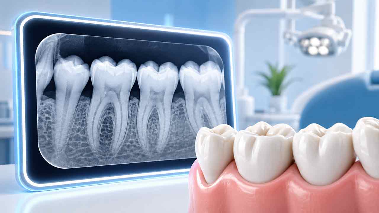
Written by: Skylar SunA dental X-ray is easy to misunderstand because the image feels definitive.There is the tooth. There is the root. There may be a dark area. Surely the picture should provide a simple yes-or-no answer.In practice, dental imaging is more useful—and more limited—than that.Radiographs can show structures that a dentist cannot directly inspect during a visual examination. They may help evaluate areas between teeth, roots, bone, developing teeth, previous dental treatment, trauma, and other clinical questions.But an X-ray does not replace the patient's history or the dentist's examination.The American Dental Association's 2026 patient-selection recommendations emphasize that dental images should be selected according to individual clinical need rather than ordered automatically on the same schedule for every patient. The recommendations call for dentists to consider medical and dental history, previous images, current clinical findings, age, disease risk, and the diagnostic information needed before selecting an imaging examination.That makes the best patient question surprisingly simple:“What question are we trying to answer with this image?”
Dental X-Rays Are Diagnostic Tools, Not Routine Photographs
A camera records what visible light can reach.Dental radiography uses X-rays to create information about internal structures and areas that cannot be inspected directly.That can make radiographs valuable for detecting or evaluating dental disease.It does not mean every patient needs every kind of image.Dentists consider the expected diagnostic benefit, the patient's clinical situation, and radiation exposure when deciding whether radiography is appropriate.
Why the Same X-Ray Is Not Right for Every Question
Different dental images are designed to show different anatomical information.A dentist evaluating possible decay between back teeth may need a different view from a dentist evaluating the root of one painful tooth.A patient undergoing an implant assessment may require information that is unnecessary during a routine cavity evaluation.That is why the phrase:“I need dental X-rays.”is incomplete.A more informative question is:Which image, for which problem, at this point in care?
Bitewing Images: Looking Between the Crowns of Teeth
Bitewing radiographs are commonly used to evaluate the crowns of upper and lower teeth together, particularly the contact areas between neighboring teeth.These areas can be difficult to inspect directly.Depending on the patient's cavity risk and clinical findings, bitewings may help a dentist evaluate decay between teeth or monitor existing findings. Imaging intervals can therefore vary between patients rather than following one universal schedule.
Periapical Images: Following the Tooth to the Root
A periapical radiograph is designed to show an individual tooth or a small group of teeth from the crown through the root and into the surrounding area.It may be useful when a dentist needs more focused information about:
- a specific tooth;
- the root;
- surrounding bone;
- previous root canal treatment;
- dental trauma;
- certain suspected endodontic problems.This type of image can provide more localized information than a broad panoramic view when the clinical question concerns one tooth or a small region.
Panoramic Images: A Wider View
A panoramic radiograph captures a broad view of the jaws and many teeth in one image.That wider field can be useful for certain questions involving:
- developing teeth;
- third molars;
- missing or additional teeth;
- jaw structures;
- dental trauma;
- some implant assessments;
- broader anatomical relationships.A panoramic image, however, is not automatically the best image for every cavity or every painful tooth.A wide image may provide useful breadth, while a focused intraoral image may provide better detail for another problem.
CBCT: Three Dimensions With a Higher Threshold
Cone-beam computed tomography, or CBCT, creates three-dimensional images of dental and maxillofacial structures.The U.S. Food and Drug Administration (FDA) explains that dental CBCT systems rotate around the patient and use a cone-shaped X-ray beam to collect data that can be reconstructed into a three-dimensional view of teeth and surrounding anatomy.That can be valuable in selected clinical situations.It is also why CBCT should not be treated as a routine “upgrade” from ordinary dental X-rays.The FDA emphasizes reducing unnecessary radiation exposure from dental CBCT and encourages clinicians to consider whether the needed information can be obtained through other appropriate imaging approaches.Depending on the clinical situation, three-dimensional imaging can involve:
- more radiation exposure than many conventional dental radiographs;
- more anatomy to interpret;
- more incidental findings;
- greater cost;
- information that may not change treatment.The best image is the one that appropriately answers the clinical question.Not the largest image.
Five Questions to Ask Before a Dental X-Ray
You do not need to understand radiographic technology to ask useful questions about dental imaging.Before an X-ray, consider asking:
- What clinical question is this image intended to answer?
- Do you already have a recent image that provides the information?
- Why is this type of image the best choice?
- Would a simpler imaging option provide the information you need?
- How will the result affect diagnosis, treatment, or monitoring?These are not refusal questions.They are informed questions.A useful answer might be very simple:
“We need a bitewing because the contact between these two teeth cannot be seen directly, and we need to check for decay.”
What Dental X-Rays Can Help Reveal
Depending on the image type and clinical context, dental radiographs may contribute information about:
- decay in areas that are difficult to see directly;
- tooth roots;
- bone surrounding teeth;
- impacted or developing teeth;
- previous root canal treatment;
- certain infections or lesions;
- dental trauma;
- implant planning;
- restoration-related concerns;
- changes compared with previous images.The wording matters:may contribute information.A radiographic appearance still needs professional interpretation in the context of the patient and examination.
What X-Rays Cannot Reliably Do Alone
An image cannot tell a dentist everything.It does not independently provide:
- the patient's pain history;
- how long a symptom lasts;
- whether a tooth hurts during chewing;
- whether cold or heat triggers discomfort;
- the feel of a suspicious tooth surface during examination;
- pulp-testing results;
- soft-tissue findings;
- every fine crack;
- whether a radiographic finding is actually responsible for the patient's symptom.Clinical context matters.A radiograph without clinical context can be easy to overinterpret.This is particularly important when patients try to obtain a diagnosis from a screenshot of an X-ray without providing the accompanying history and examination findings.
Why a Dark Area Is Not a Diagnosis
Patients often notice dark regions on dental radiographs.Dental radiographs are created by differences in how tissues and materials interact with X-rays. Normal anatomical spaces, tooth structures, restorative materials, disease processes, and image geometry can therefore appear differently on the image.The word “dark” therefore does not mean “cavity.”Likewise, a light area does not automatically mean “healthy.”A dentist interprets factors such as:
- location;
- shape;
- boundaries;
- tooth anatomy;
- surrounding structures;
- clinical findings;
- previous images.If an area concerns you, a useful question is:
“What anatomical structure am I looking at, and what makes this appearance normal or abnormal?”That creates a more informative conversation than trying to name a disease from the image yourself.
The Value of Previous X-Rays
One of the strongest uses of dental imaging is comparison over time.A single image shows one moment.Two appropriately comparable images may help a dentist determine whether something appears stable or whether it has changed.The ADA's patient-selection and radiation-safety guidance encourages dental professionals to consider previously acquired images when they can provide useful diagnostic information.If you change dentists, transferring relevant dental records and radiographs may therefore be helpful.A new dentist may still decide that additional imaging is clinically necessary, but access to previous images can provide useful history and may help avoid unnecessary duplication.
There Is No Universal “Every Six Months” X-Ray Rule
Many patients assume dental X-rays should automatically be taken at every cleaning.Others believe X-rays should be avoided unless they have severe pain.Neither rule reflects individualized dental imaging.The ADA's 2026 patient-selection recommendations emphasize that decisions about dental radiography should account for factors such as:
- medical and dental history;
- previous radiographs;
- current clinical findings;
- age and dental development;
- cavity risk;
- periodontal findings;
- treatment needs;
- the specific diagnostic question.The appropriate interval can therefore differ among patients.It can also change over time for the same person.Someone with active dental disease may need a different monitoring approach from someone whose risk and clinical findings remain consistently low.
Radiation Risk Should Be Minimized, Not Dramatized
Dental radiography uses ionizing radiation.That fact should neither be hidden nor sensationalized.The goal of responsible imaging is to obtain clinically useful information while avoiding unnecessary exposure.Modern radiation-safety practices can include:
- selecting images only when clinically justified;
- using appropriate digital imaging techniques;
- restricting the X-ray beam to the area that needs assessment;
- positioning the patient correctly;
- avoiding unnecessary repeat images;
- using previous images when they remain diagnostically useful.The principle is not:“More imaging is better.”It is also not:“Any radiation means imaging should be avoided.”The decision should balance diagnostic value with appropriate radiation protection.
Why Routine Lead Aprons Have Changed
This is one area where a patient's previous dental experience may differ from current recommendations.In its 2024 radiation-safety recommendations, the ADA stated that routine use of lead abdominal aprons and thyroid collars is no longer recommended during dental radiography when modern imaging practices are followed.The ADA explained that current digital imaging technology and restricting the X-ray beam to the area being examined provide effective ways to reduce unnecessary exposure.Protective shielding can also interfere with the primary X-ray beam in some situations. If that blocks part of the needed image, another radiograph may have to be taken.The recommendations instead emphasize:
- justified image selection;
- digital imaging;
- appropriate beam restriction;
- proper patient positioning;
- avoiding unnecessary repeat exposures.Dental practices must still comply with applicable federal, state, and local regulations.The change does not mean radiation protection is no longer important.It reflects a change in how modern dental radiation protection is approached.
Why CBCT Is Not Automatically “More Accurate”
Three-dimensional imaging can provide information that two-dimensional radiographs cannot.But more dimensions do not automatically make an examination more appropriate for a particular clinical question.A focused two-dimensional image may provide all the information needed in one situation, while CBCT may be useful when additional three-dimensional detail is genuinely necessary.More imaging capability is not the same thing as more appropriate imaging.
Dental X-Rays and Tooth Decay
Radiographs can be particularly useful when decay is located in areas that are difficult to inspect directly, especially between teeth.But radiographic detection also has limitations.Very early mineral changes may not always create an obvious radiographic appearance.A dentist can combine imaging with visual examination, cavity history, tooth anatomy, current risk factors, and previous radiographs.This explains why an early enamel change can sometimes be discussed clinically even when a patient cannot see an obvious “hole” on an X-ray.A dental radiograph is not a binary cavity scanner.
Dental X-Rays and Root Canal Problems
When a tooth is painful, a dentist may use periapical radiographs to evaluate the root and surrounding structures.Endodontic diagnosis still relies on more than radiography. A dentist may consider:
- symptoms;
- clinical examination;
- pulp testing;
- periodontal findings;
- radiographic appearance.A tooth should not be assigned root canal treatment solely because one area appears dark on an image without appropriate clinical correlation.The radiograph contributes evidence.It does not independently determine treatment.
Imaging After Dental Trauma
A tooth that has been hit, displaced, fractured, or otherwise injured may need radiographic evaluation.Imaging can help a dentist investigate structures that cannot be fully assessed by looking at the tooth alone.Follow-up may also matter because the condition of an injured tooth can change over time.A tooth that looks normal immediately after an accident does not necessarily eliminate the need for further assessment when the type of injury or later symptoms warrant it.The appropriate imaging method and follow-up schedule depend on:
- the type of trauma;
- the tooth involved;
- the patient's age;
- root development;
- clinical findings;
- symptoms;
- changes over time.The dentist can determine which images, if any, are appropriate for the individual injury.
Keep an X-Ray Comparison Log
If a dentist is monitoring one tooth or one area over time, keeping a short record can make future conversations easier.
This can be particularly useful when a dentist says:
“We're going to watch this area.”“Watch” should have a purpose.You can ask what finding is being monitored and what type of change would matter.The log helps preserve that information between visits without asking patients to interpret the X-rays themselves.
A Good Image Does Not Guarantee a Good Diagnosis
A technically excellent dental image can still be misunderstood when it is separated from the patient and clinical context.High image quality improves the information available to the dentist, but it does not create diagnostic certainty by itself.This is also why an AI-highlighted X-ray, a screenshot sent online, or a single image viewed remotely should not automatically replace a complete dental evaluation when one is needed.The image is evidence. It is not the entire case.
Bottom Line
Dental X-rays are most useful when they answer a specific clinical question that cannot be answered as well through examination alone.They are not photographs that every patient automatically needs on the same schedule.They are not perfect scanners that identify every dental problem.And a three-dimensional scan is not automatically superior to a focused two-dimensional image.The American Dental Association's 2026 patient-selection recommendations emphasize using dental imaging according to individual clinical need. Its 2024 radiation-safety recommendations likewise focus on justified imaging, modern radiation-protection practices, and avoiding unnecessary exposures.The FDA also encourages appropriate use of dental CBCT and reducing unnecessary radiation exposure.The most useful question a patient can ask is not simply:“Do I need X-rays every year?”It is:“What information do you need from this image, and how will it affect my care?”That keeps dental imaging where it belongs:as a targeted diagnostic tool used to support better dental decisions.
Authoritative Sources
- American Dental Association — 2026 Dental X-Ray Patient Selection Recommendations
- American Dental Association — 2024 Radiography Safety Recommendations
- U.S. Food and Drug Administration — Dental Cone-Beam Computed Tomography
About the Author
Skylar Sun writes consumer-focused dental health explainers designed to make everyday dental topics easier to understand.Skylar's research approach relies on publicly available guidance from established professional and government health organizations, including the American Dental Association (ADA), the U.S. Food and Drug Administration (FDA), and other authoritative dental-health sources relevant to each topic.Articles are developed with an emphasis on source transparency, careful interpretation of health information, practical reader questions, and clear distinctions between general education and individualized dental care.